The Philosophy: “Defects are the Active Sites”
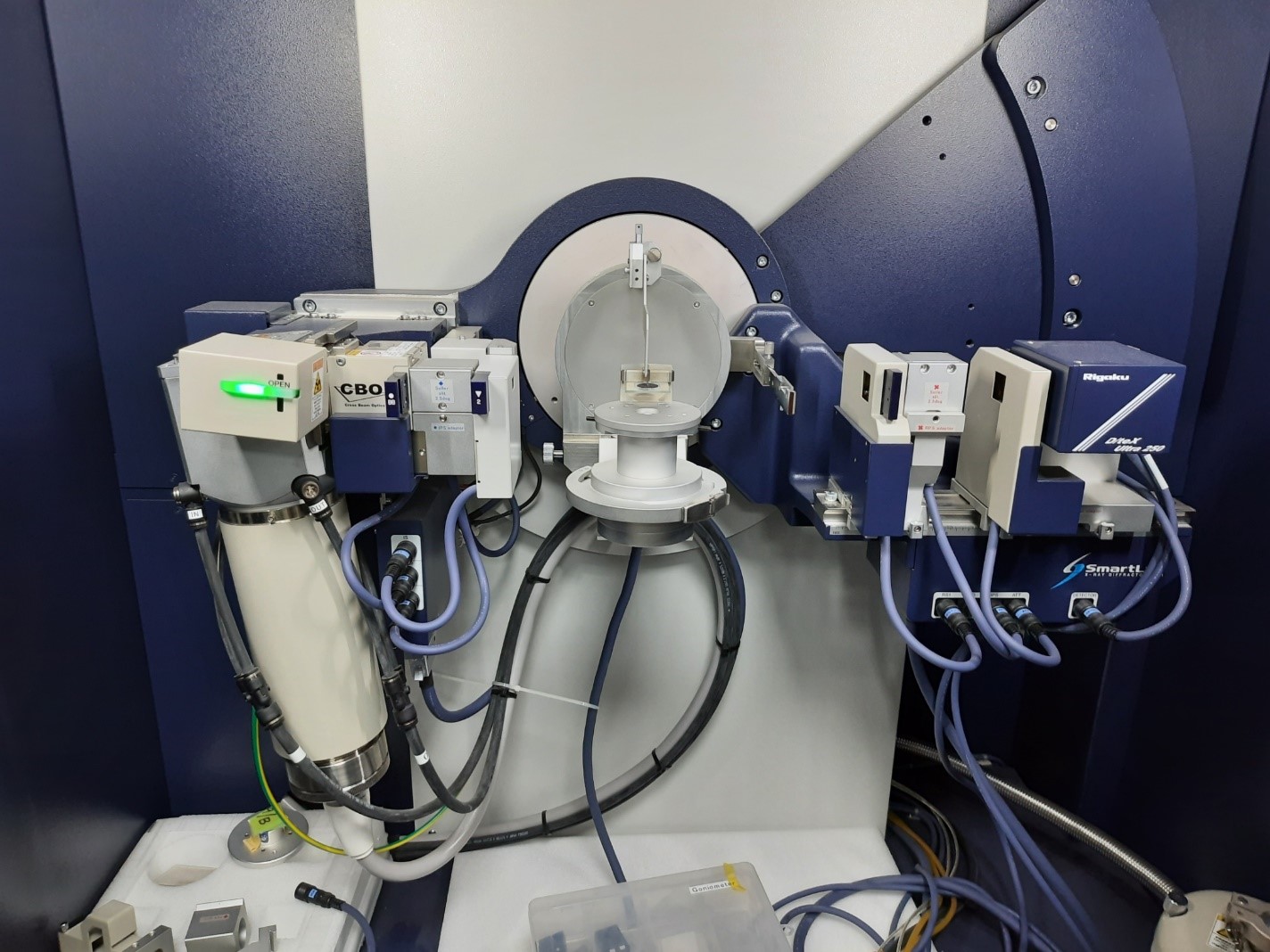
In classical materials science, a perfect crystal is the ideal. In energy storage and catalysis, however, imperfection is the key. Grain boundaries, stacking faults, and lattice strain which exist on the Ångström scale are often where the chemistry happens. Bulk characterization (like XRD) gives an average picture, but it cannot explain why two chemically identical samples perform differently.
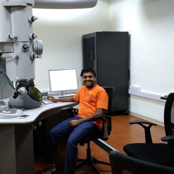
My Expertise: Seeing the Invisible I utilize Transmission Electron Microscopy (TEM) not just to image materials, but to perform forensic analysis on their atomic structure. Moving beyond bulk averages (XRD), I use direct electron imaging to correlate local disorder with macroscopic device performance.
Technique 1: Lattice Fringe Analysis (Thesis Work)
Validating the success of a synthesis protocol requires proving phase purity at the single-particle level.
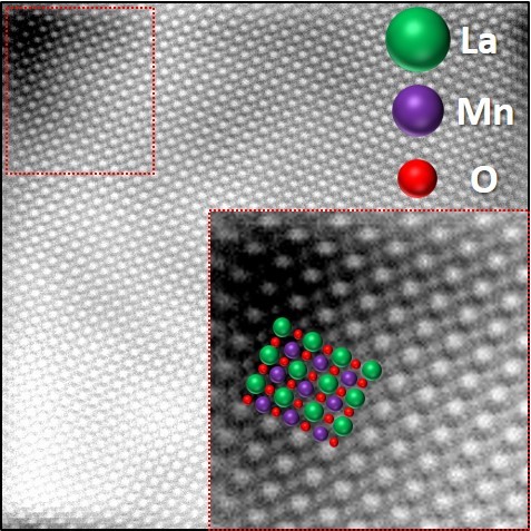
- Method: High-Resolution TEM (HRTEM) on synthesized Lanthanum Perovskite Nanorods.
- Analysis: * FFT (Fast Fourier Transform): Converted real-space lattice images into reciprocal space patterns to confirm single-crystalline nature.
- d-spacing: Precisely measured interplanar distances (e.g., ~$0.27$ nm for the (110) plane) to distinguish between competitive orthorhombic and rhombohedral phases.
Technique 2: “Turbostratic” Disorder in NiFe-LDH
From recent work on Battery-Type Supercapacitors.
The Anomaly: We synthesized NiFe-Layered Double Hydroxides (LDH) for supercapacitors, but specific synthesis batches showed anomalously high capacity, defying theoretical predictions for pristine crystals. The Hypothesis: We suspected that “stacking faults”—errors in the layering sequence—were creating extra active sites.
The Discovery: I utilized High-Resolution TEM (HRTEM) to image the lattice fringes directly.
- Technique: Bright Field Imaging & Selected Area Electron Diffraction (SAED).
- Result: The imaging revealed distinct “turbostratic” disorder (rotational misalignment) between the hydroxide layers, which facilitated faster ion diffusion.
- The Mechanism: This “messy” stacking expanded the interlayer gallery height.
- The Impact: This reduced the diffusion barrier for electrolyte ions, effectively turning a standard battery material into a high-rate supercapacitor electrode.
Visual Methodology
📜 Source & Citation
Note: Portions of this methodology are derived from the author's doctoral work. Please cite as follows:
Kotha, V. (2022). Tailoring Transition Metal Perovskite Oxides via Low-Temperature Hydrothermal Routes as Potential Candidates for Catalytic Applications [Doctoral dissertation, IIT Bombay].